Genetic manipulation is a standard research approach for cardiac hypertrophy and heart failure. We use adenoviral transfection for overexpression, dominant inhibition, or RNA suppression in adult, neonatal, or immortalized cardiac myocytes to generate mechanistic data, and genetic manipulation (overexpression and/or gene ablation) in mouse hearts for functional studies.
Recently, we have developed techniques for cardiac-specific gene manipulation and phenotype analysis in Drosophila melanogaster, the fruit fly, to bridge the time and resource gap between adenovirally transfected cardiac myocytes and genetic mouse models. Drosophila studies are facilitated by a genetic tool kit that we use to rapidly manipulate multiple genes in a cardiac specific manner. Fly hearts are sufficiently like those of mammals to permit analysis of cardiac structure and function using analagous phenotyping techniques. Fly hearts are also useful for live cell studies. Ongoing studies are focused on the effects of mitochondrial fusion and fission on the heart.
Non-invasive assessment of cardiac function in D. melanogaster
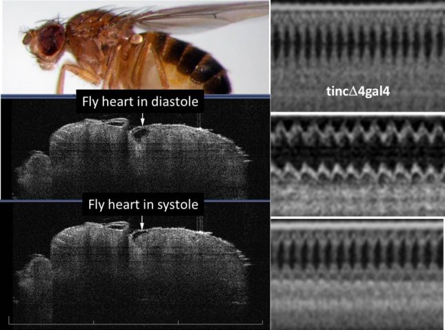
Optical coherence tomography imaging of functioning heart tubes in intact, unanesthetized fruit flies using a Michelson Diagnostics 1300 nm multi-beam EX1301 OCT microscope (greater than 10 um resolution). Two-dimensional images (left panels) are post-processed to achieve b-mode tracings analogous to mouse/human m-mode echocardiography studies (right panels). Top right tracing is normal control (tincD4gal4) heart. Middle right tracing is dilated hypocontractile cardiomyopathic heart that was rescued genetically (bottom tracing).
Real time imaging of contracting D. melanogaster heart tube
Green fluorescent protein was expressed specifically in cardiac myocytes, and cardiac contractions imaged using a GFP ultraviolet fluorescence microscope.
Live-cell confocal imaging of cardiac myocyte mitochondria in D.melanogaster
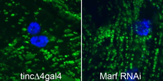
Flies were generated that express mitochondrial-localized GFP only in the heart, which is useful to assess mitochondrial morphology, distribution, and trafficking in isolated D. melanogaster heart tubes in short-term culture. Green GFP labels mitochondria, blue Hoechst dye labels nuclei.