Delineation of a Molecular Mechanism for Programmed Cardiac Myocyte Death After Myocardial Infarction
Programmed cardiac myocyte death (apoptosis) increases cardiac damage late after myocardial infarction by extending the infarct and through loss of cardiac myocytes in non-infarcted myocardium. The programmed cell death gene BNip3 was reported by several groups as upregulated in ischemic hearts. We determined the consequences of BNip3 upregulation in mice by forcing its expression through cardiac-specific transgenesis, and by knocking the BNip3 gene out of the mouse genome.
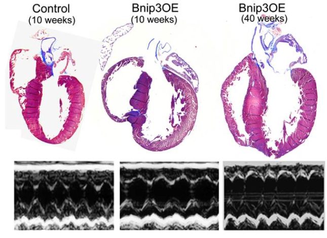
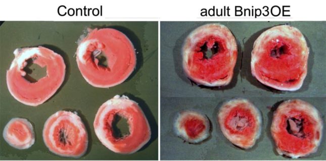
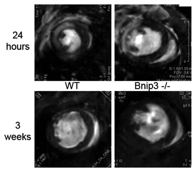
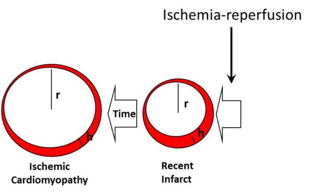
BNip3 causes heart failure from programmed cardiac myocyte death. The heart on the left is a normal control. Below is its M-mode echocardiogram, showing normal left ventricular contractions. The middle heart is from a 10 week old (young adult) mouse expressing BNip3 in the heart. The heart is enlarged and the echocardiographic ventricular shortening is reduced. The right heart is a 40 week old BNip3 transgenic mouse (fully mature adult). This heart is also enlarged with dilated ventricular chambers. The echocardiogram reveals severe cardiomyopathy.BNip3 increases myocardial infarction size. Mice underwent experimental myocardial infarction by surgically ligating the left anterior descending coronary artery. After 24 hours, the hearts are removed, sectioned, and stained to assess infarct size. Healthy myocardium stains red and infarcted myocardium appears white. The BNip3 hearts show larger, more extensive, and diffuse infarcts due to programmed cell death.BNip3 gene knockout mice are protected from heart failure after myocardial infarction. These are cine magnetic resonance images (cMRI) of the same normal (WT, wild-type, left) and BNip3 gene knockout (Bnip3-/-, right) mouse hearts one day (top) and 3 weeks (bottom) after myocardial infarction. The infarctions are visualized in 24 hour studies by gadolinium staining that appears white (areas between 9 and 12 o’clock) within the dark myocardium. Blood within the left ventricles appears bright white in these images and swirls as it is ejected from the heart. After three weeks the infarct size (area of the heart that doesn’t move) is smaller in BNip3 knockout hearts.Schematic depiction of the role of BNip3 and programmed cell death (apoptosis) in cardiomyopathy after myocardial infarction/ischemia-reperfusion.